Central Canal Stenosis Grading: What Mild, Moderate, and Severe Mean on MRI
Central canal stenosis means the main tunnel for the spinal cord or nerve roots is narrowed, but the grade — mild, moderate, or severe — only describes the MRI appearance and does not automatically predict pain level, disability, or need for surgery.
An MRI, or magnetic resonance imaging scan, gives detailed pictures of the spine. It can show narrowing, disc problems, arthritis, and pressure on nerves. But the MRI report is only one part of the story.
In my practice, I tell patients that the grade is a starting point, not the final diagnosis. The key question is not just, “How narrow does it look?” The better question is, “Is that narrowing affecting the spinal cord or nerve roots in a way that matches your symptoms?”
What Is Central Canal Stenosis?
The central canal is the main passageway through the spine.
In the neck, also called the cervical spine, the central canal contains the spinal cord. The spinal cord is the main bundle of nerves that carries signals between your brain and body.
In the lower back, also called the lumbar spine, the spinal cord has usually ended. The canal contains nerve roots, which are smaller nerves that travel from the spine into the legs. These nerve roots are called the cauda equina, which means “horse’s tail,” because of how they look.
Stenosis means narrowing.
So, central canal stenosis means the main spinal tunnel is narrowed. The key question is not just “how narrow does it look?” but “is that narrowing affecting the spinal cord or nerve roots in a way that matches the patient’s symptoms?”
Central canal stenosis is not the same as every other type of spine narrowing.
Central canal vs. foraminal vs. lateral recess stenosis
There are three common places where narrowing can happen:
- Central canal stenosis: narrowing of the main spinal canal.
- Foraminal stenosis: narrowing of the side exits where nerves leave the spine. These side exits are called foramina.
- Lateral recess stenosis: narrowing of the pathway just before a nerve exits the spine.
These can overlap. A person may have central stenosis, foraminal stenosis, lateral recess stenosis, or a mix of all three.
To learn more about side-exit narrowing, see Neural Foraminal Narrowing: What Mild, Moderate, and Severe Mean.
For narrowing just before the nerve exits, see Lateral Recess Stenosis: The Stenosis Patients Don’t Know They Have.
What Do Mild, Moderate, and Severe Central Canal Stenosis Mean?
Radiologists often use the words mild, moderate, and severe to describe how much narrowing they see on MRI.
These words are helpful. But they are not exact treatment instructions.
What I look for on MRI is whether there is still room around the nerves or spinal cord, not just the single word used in the report.
Mild central canal stenosis
Mild central canal stenosis usually means the canal is smaller than normal, but the MRI does not show major crowding of the spinal cord or nerve roots.
There is often still enough space around the nerves or spinal cord.
Mild stenosis can happen from early age-related spine changes, such as:
- A disc bulge, meaning the soft cushion between the bones pushes outward.
- Facet enlargement, meaning the small joints in the back of the spine become arthritic and bigger.
- Ligament thickening, meaning a band of tissue near the canal gets thicker and takes up more space.
Mild stenosis may or may not be related to your symptoms. Sometimes it is just one finding among several changes on the MRI.
Moderate central canal stenosis
Moderate central canal stenosis means the narrowing is more noticeable.
The normal fluid space around the spinal cord or nerve roots may be reduced. This fluid is called cerebrospinal fluid, or CSF. It cushions the nerves and spinal cord.
In the lumbar spine, moderate stenosis may make the nerve roots look more crowded. In the cervical spine, the spinal cord may have less room.
But there are two separate questions in the neck:
- Is the spinal cord compressed?
- Is there cord signal change, meaning an abnormal bright or dark area inside the spinal cord on MRI that may suggest stress or injury?
“Moderate central stenosis” does not automatically mean surgery. It means the narrowing is significant enough that your doctor should compare the MRI with your symptoms, walking tolerance, neurologic exam, and the exact spinal level involved.
A neurologic exam is a physical exam that checks nerve and spinal cord function. It may include strength, feeling, reflexes, balance, and walking.
Severe central canal stenosis
Severe central canal stenosis means there is marked narrowing of the canal.
In the lower back, the nerve roots may be tightly crowded. In the neck, the spinal cord may be flattened or compressed.
Compression means pressure on a nerve or the spinal cord.
Severe central canal stenosis is a finding doctors take seriously, but the MRI grade alone still does not tell the whole story. The most important question is whether the stenosis is causing nerve or spinal cord dysfunction.
Dysfunction means the nerve or spinal cord is not working normally.
Severe cervical stenosis needs special attention for signs of myelopathy. Myelopathy means spinal cord dysfunction. It can affect balance, hand use, walking, strength, or feeling.
Severe lumbar stenosis needs to be matched with leg symptoms, walking limits, and nerve findings on exam.
How Radiologists Grade Central Canal Stenosis
A radiologist is a doctor who reads imaging studies, such as MRI scans.
Radiologists commonly use terms like mild, moderate, and severe. But there is not one universal grading system used in every MRI report.
Grading may be based on:
- How much space remains in the canal.
- Whether the spinal fluid around the nerves or cord is reduced.
- Whether the nerve roots are crowded.
- Whether the spinal cord is flattened.
- Whether there is abnormal spinal cord signal.
Some research grading systems look closely at the shape of the nerve sac and how crowded the nerve roots are. But many everyday MRI reports use more general description words.
The same MRI can sometimes be described differently by different readers. One radiologist may say “moderate to severe.” Another may say “severe.” This does not always mean the condition changed. It may reflect judgment in how the images are described.
Why the exact MRI images matter
The written report is useful, but it is a summary.
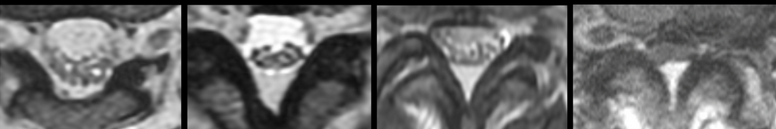
A spine surgeon often wants to see the actual MRI images, not just the report. The axial images are especially important. Axial images are cross-section views, like looking at a slice through the spine.
What I look for on MRI is not just the word “moderate” or “severe,” but the actual space available for the nerves or spinal cord at each level.
For more help with MRI wording, see How to Read Your Spine MRI Report.
Lumbar vs. Cervical Central Canal Stenosis: Why Location Matters
The finding matters most when I know where it is: stenosis in the neck and stenosis in the lower back can behave very differently.
Lumbar central canal stenosis
Lumbar central canal stenosis occurs in the lower back.
It usually affects the cauda equina nerve roots that travel into the legs.
A classic symptom pattern is called neurogenic claudication. This means leg symptoms caused by nerve crowding in the spine, often worse with standing or walking.
Lumbar stenosis may cause:
- Leg pain.
- Leg heaviness.
- Numbness.
- Tingling.
- Symptoms that get worse with standing or walking.
- Relief with sitting or leaning forward.
Back pain can happen with lumbar stenosis, but back pain alone does not prove the stenosis is the pain generator.
To learn more, see Lumbar Spinal Stenosis: A Plain-Language Guide for Patients.
Cervical central canal stenosis
Cervical central canal stenosis occurs in the neck.
This location matters because the neck contains the spinal cord. If cervical stenosis affects the spinal cord, doctors worry about cervical myelopathy.
Symptoms of spinal cord involvement may include:
- Balance trouble.
- Hand clumsiness.
- Difficulty with buttons.
- Changes in handwriting.
- Trouble with fine motor tasks, meaning small hand movements.
- Weakness.
- Numbness or tingling in the arms or legs.
- Changes in walking.
For more detail, see Cervical Spinal Stenosis & Cervical Myelopathy.
Does Central Canal Stenosis Always Cause Symptoms?
No.
MRI findings and symptoms do not always match perfectly. Some people have central canal stenosis on MRI and few symptoms. Some people have pain from another structure, even when stenosis is also present.
Other possible pain sources include:
- Disc herniation, meaning disc material pushes out of place.
- Foraminal stenosis.
- Lateral recess stenosis.
- Facet arthritis, meaning arthritis in the small joints in the back of the spine.
- Sacroiliac joint pain, meaning pain from the joint between the spine and pelvis.
- Hip, knee, nerve, or blood vessel conditions.
MRI findings are clues, not the whole diagnosis. The finding matters most when the level and side of narrowing match the patient’s symptoms and exam.
The MRI has to match the story. If the symptoms, exam, and imaging all point to the same level, the finding becomes much more meaningful.
For leg pain that may come from a spinal nerve, see Sciatica: Causes, Diagnosis, and the Treatment Path.
For arthritis in the small spine joints, see Facet Arthropathy and Facet Joint Hypertrophy.
Common Causes of Central Canal Stenosis on MRI
Central canal stenosis often comes from a mix of age-related spine changes. These changes can take up space inside the canal.
Disc bulge or disc herniation
A disc is the cushion between two spine bones.
A disc bulge means the disc extends outward around part or all of its edge. A disc herniation means a more focused area of disc material pushes out of place.
Disc material can push backward into the central canal. Central or paracentral disc herniations may contribute to canal narrowing. Paracentral means slightly off to one side of the center.
Learn more here:
- Disc Bulge vs. Protrusion vs. Extrusion vs. Sequestration
- Foraminal vs. Paracentral vs. Central Disc Herniations
Ligamentum flavum hypertrophy
The ligamentum flavum is a ligament in the back part of the spinal canal. A ligament is a strong band of tissue that connects bones.
Hypertrophy means thickening or enlargement.
Ligamentum flavum hypertrophy means this ligament has thickened and may buckle inward. This is a common contributor to lumbar stenosis.
See Ligamentum Flavum Hypertrophy: Why Your Ligament Is Making Your Canal Smaller.
Facet joint hypertrophy
The facet joints are small joints in the back of the spine. They help guide motion.
Facet joint hypertrophy means these joints have enlarged, often due to arthritis. This can narrow the canal, the lateral recess, or the foramen.
See Facet Arthropathy and Facet Joint Hypertrophy.
Spondylolisthesis
Spondylolisthesis means one spine bone has slipped forward or backward compared with the bone next to it.
A slipped vertebra can narrow the central canal. A vertebra is one of the bones of the spine.
The amount of slip and whether the bones move too much can affect treatment decisions.
See Spondylolisthesis: When the Bones Slip.
When Central Canal Stenosis Is More Concerning
Central canal stenosis deserves closer attention when it is linked with signs that nerves or the spinal cord are not working well.
More concerning findings include:
- Progressive weakness.
- Worsening numbness.
- Trouble walking.
- Balance problems.
- Hand clumsiness in cervical stenosis.
- Spinal cord compression.
- Abnormal spinal cord signal change.
- Bowel or bladder dysfunction.
- Saddle anesthesia.
Saddle anesthesia means numbness in the groin, inner thighs, buttocks, or the area that would touch a saddle.
Seek urgent medical care now if you have new loss of bowel or bladder control, numbness in the groin or saddle area, rapidly worsening leg weakness, severe new difficulty walking, or symptoms suggesting spinal cord dysfunction such as new balance problems, hand clumsiness, or progressive weakness. SpineClarity’s written review service is not for emergencies.
One emergency to know is cauda equina syndrome. This means severe pressure on the bundle of nerve roots in the lower spine. It can affect bladder, bowel, sexual function, leg strength, and feeling.
Read more here: Cauda Equina Syndrome: The Spine Emergency Patients Need to Recognize.
Does Moderate or Severe Central Canal Stenosis Mean I Need Surgery?
Not necessarily.
Surgery is not based on the MRI word alone. It is based on whether the stenosis is causing a problem that is significant enough — and anatomically clear enough — that decompression is likely to help.
Decompression means surgery to create more space for the nerves or spinal cord.
Treatment decisions depend on:
- Severity of symptoms.
- How long symptoms have been present.
- Neurologic deficits, meaning weakness, numbness, reflex changes, or spinal cord signs.
- Walking tolerance.
- Cervical vs. lumbar location.
- Whether the spinal cord is compressed.
- Whether there is spinal cord signal change.
- Response to non-surgical care.
Many people start with non-surgical treatment if there are no urgent neurologic problems. Non-surgical care may include activity changes, physical therapy, medicines, or injections, depending on the situation.
Surgery is more likely to be discussed when symptoms are disabling, progressive, or when neurologic function is threatened.
I do not recommend treatment based on the word “severe” alone. I want to know what the patient can and cannot do, whether there are neurologic deficits, and whether the anatomy explains the symptoms.
How a Spine Surgeon Interprets Central Canal Stenosis on MRI
A spine surgeon does not just read the label. The goal is to connect the MRI anatomy with the patient’s story and exam.
In my practice, the most important step is matching the MRI to the patient. A report that says “severe stenosis” may be very important — but only in the context of where it is, what structures are compressed, and what the patient is actually experiencing.
A practical checklist includes:
- Which level is narrowed?
- Is it cervical, thoracic, or lumbar?
- Is there spinal cord compression?
- Is there cord signal change?
- Are the nerve roots crowded?
- Is the stenosis central, foraminal, lateral recess, or mixed?
- Is the stenosis caused by disc, bone, ligament, slip, cyst, or a combination?
- Does the imaging match the patient’s symptoms?
- Are there neurologic deficits?
- Are there red flags?
The thoracic spine is the mid-back area. Thoracic central stenosis is less common than cervical or lumbar stenosis, but it can matter because the spinal cord also runs through this area.
A cyst is a fluid-filled sac. In the spine, some cysts can come from arthritic joints and narrow the canal.
When a Written MRI Review Can Help
If your MRI report says “moderate” or “severe central canal stenosis” and you are not sure what it means for your symptoms, SpineClarity can help you understand the report in plain language.
SpineClarity offers a written MRI/case review from a board-certified spine surgeon. You can upload your symptoms, MRI report, and relevant records, and receive a plain-language written interpretation with a suggested next-step category. This is not emergency care and does not replace an in-person physician relationship.
Frequently Asked Questions
What does central canal stenosis mean on MRI?
Central canal stenosis means the main tunnel through the spine is narrower than expected.
In the neck, that tunnel contains the spinal cord. In the lower back, it contains the cauda equina nerve roots that travel into the legs.
What is the difference between mild, moderate, and severe central canal stenosis?
Mild means the canal is narrowed, but there is usually still good space around the nerves or spinal cord.
Moderate means the narrowing is more noticeable. The fluid space may be reduced, and nerves may look more crowded.
Severe means marked narrowing. Nerve roots may be crowded, or the spinal cord may be compressed.
These grades describe the MRI appearance. They do not decide treatment by themselves.
Is moderate central stenosis serious?
Moderate central stenosis can be important, but it does not automatically mean surgery.
It matters more if the location of narrowing matches your symptoms and exam. It also matters whether it is in the neck or lower back.
Moderate cervical stenosis may need closer attention if there are signs of spinal cord involvement.
Does severe central canal stenosis mean I need surgery?
Not always.
Severe stenosis is taken seriously, but treatment depends on the full picture. That includes symptoms, walking ability, neurologic findings, spinal cord compression, cord signal change, and response to non-surgical care.
Can central canal stenosis cause leg pain or sciatica?
Yes, lumbar central canal stenosis can cause leg pain, heaviness, numbness, or tingling.
This often gets worse with standing or walking and improves with sitting or leaning forward.
Sciatica means pain that travels down the leg from irritation of a spinal nerve. Central stenosis can contribute to leg symptoms, but foraminal stenosis, lateral recess stenosis, and disc herniation can also cause similar pain.
Can central canal stenosis cause numbness or weakness?
Yes.
Central canal stenosis can cause numbness, tingling, or weakness if it affects nerve roots or the spinal cord.
Progressive weakness or worsening numbness deserves prompt attention, especially if it affects walking, balance, or hand function.
Is cervical central canal stenosis more dangerous than lumbar stenosis?
Cervical stenosis can be more concerning when it affects the spinal cord.
Lumbar stenosis usually affects nerve roots in the lower back. Cervical stenosis can affect the spinal cord itself, which can lead to myelopathy.
That does not mean every cervical stenosis finding is an emergency. It means symptoms and cord findings matter.
What does it mean if my MRI says there is spinal cord compression?
Spinal cord compression means the cord is being pressed or flattened.
This can be important, especially if you have balance problems, hand clumsiness, walking changes, weakness, or numbness.
The degree of compression, the exact level, and whether there is cord signal change all matter.
What does cord signal change mean with stenosis?
Cord signal change means the spinal cord looks abnormal on MRI in the area of compression.
It may suggest stress or injury to the spinal cord. It does not always mean the same thing for every person, but doctors usually take it seriously in the setting of cervical stenosis or myelopathy symptoms.
Can I have central canal stenosis without symptoms?
Yes.
Some people have central canal stenosis or other age-related MRI findings with little or no symptoms.
That is why the MRI must be matched with the symptom pattern and physical exam.
What symptoms should make me seek urgent care?
Seek urgent medical care now for:
- New loss of bladder or bowel control.
- New trouble urinating.
- Numbness in the groin or saddle area.
- Rapidly worsening leg weakness.
- Severe new difficulty walking.
- New balance problems.
- New hand clumsiness.
- Progressive arm or leg weakness.
- Worsening symptoms after trauma.
SpineClarity’s written review service is not for emergencies.
Should I get a second opinion on my MRI report?
A second opinion can be helpful when the report says “moderate” or “severe” stenosis and you are unsure what it means.
It can also help when symptoms and MRI findings do not seem to match, when surgery has been discussed, or when the report mentions spinal cord compression or cord signal change.
A written MRI review can help explain the imaging in plain language. It does not replace an in-person exam or emergency care.
Related Articles
References
Boden SD, Davis DO, Dina TS, Patronas NJ, Wiesel SW. Abnormal magnetic-resonance scans of the lumbar spine in asymptomatic subjects: A prospective investigation. Journal of Bone and Joint Surgery American Volume. 1990;72(3):403-408.
Brinjikji W, Luetmer PH, Comstock B, et al. Systematic literature review of imaging features of spinal degeneration in asymptomatic populations. AJNR American Journal of Neuroradiology. 2015;36(4):811-816. doi:10.3174/ajnr.A4173
Davies BM, Mowforth OD, Smith EK, Kotter MRN. Degenerative cervical myelopathy. BMJ. 2018;360:k186. doi:10.1136/bmj.k186
Fehlings MG, Tetreault LA, Riew KD, et al. A clinical practice guideline for the management of patients with degenerative cervical myelopathy. Global Spine Journal. 2017;7(3 Suppl):70S-83S. doi:10.1177/2192568217701914
Fraser S, Roberts L, Murphy E. Cauda equina syndrome: A literature review of its definition and clinical presentation. Archives of Physical Medicine and Rehabilitation. 2009;90(11):1964-1968. doi:10.1016/j.apmr.2009.03.021
Lee GY, Lee JW, Choi HS, Oh KJ, Kang HS. A new grading system of lumbar central canal stenosis on MRI: An easy and reliable method. AJR American Journal of Roentgenology. 2011;196(5). doi:10.2214/AJR.10.5560
Lurie J, Tomkins-Lane C. Management of lumbar spinal stenosis. BMJ. 2016;352:h6234. doi:10.1136/bmj.h6234
Munakomi S, Foris LA, Varacallo M. Spinal stenosis and neurogenic claudication. In: StatPearls. Treasure Island, FL: StatPearls Publishing. NCBI Bookshelf.
North American Spine Society. Evidence-Based Clinical Guidelines for Multidisciplinary Spine Care: Diagnosis and Treatment of Degenerative Lumbar Spinal Stenosis. Burr Ridge, IL: North American Spine Society; 2011.
Nouri A, Tetreault L, Singh A, Karadimas SK, Fehlings MG. Degenerative cervical myelopathy: Epidemiology, genetics, and pathogenesis. Spine. 2015;40(12):E675-E693. doi:10.1097/BRS.0000000000000913
Schizas C, Theumann N, Burn A, et al. Qualitative grading of severity of lumbar spinal stenosis based on the morphology of the dural sac on magnetic resonance images. Spine. 2010;35(21):1919-1924. doi:10.1097/BRS.0b013e3181d359bd
Spector LR, Madigan L, Rhyne A, et al. Cauda equina and conus medullaris syndromes. In: StatPearls. Treasure Island, FL: StatPearls Publishing. NCBI Bookshelf.
Tetreault LA, Dettori JR, Wilson JR, et al. Systematic review of magnetic resonance imaging characteristics that affect treatment decision making and predict clinical outcome in patients with cervical spondylotic myelopathy. Spine. 2013;38(22 Suppl 1):S89-S110. doi:10.1097/BRS.0b013e3182a7eae0
Weinstein JN, Tosteson TD, Lurie JD, et al. Surgical versus nonsurgical therapy for lumbar spinal stenosis. New England Journal of Medicine. 2008;358(8):794-810. doi:10.1056/NEJMoa0707136