Lateral Recess Stenosis on MRI: What It Means and When It Matters
Lateral recess stenosis means that one of the small side channels inside the spinal canal has become narrowed, sometimes crowding the nerve root that travels through that area toward the leg.
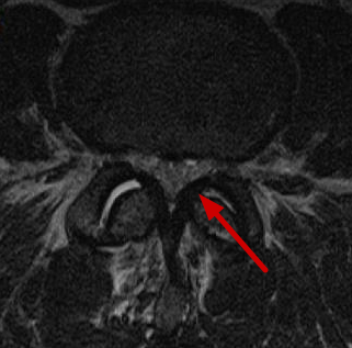
MRI means magnetic resonance imaging. It is a scan that shows the discs, nerves, joints, and soft tissues of the spine. Stenosis means narrowing. So if your lumbar MRI report mentions “lateral recess stenosis” or “subarticular recess narrowing,” it is describing a narrow space near a nerve.
This finding can matter. It can also be present without causing symptoms. The key question is whether the MRI finding matches your leg pain, numbness, tingling, weakness, and exam findings.
What Is Lateral Recess Stenosis?
The spinal canal is the main tunnel inside your spine. It holds the nerve structures that travel down toward your legs.
The lateral recess is a small side passage inside that canal. It sits near the path of a nerve root before the nerve leaves the spine through the foramen. A nerve root is a branch of nerve that leaves the spine and travels into the leg. The foramen is the small exit opening where the nerve leaves the spine.
Lateral recess stenosis means this side passage has narrowed.
This is also called subarticular recess stenosis. “Subarticular” means under or near the facet joint, which is one of the small joints in the back of the spine.
This finding is most often discussed in the lumbar spine. Lumbar means the lower back.
A simple way to picture it:
The spinal canal is like a hallway. The lateral recess is a side lane where a nerve root passes before leaving the spine. If that side lane narrows, the nerve can become crowded.
In my practice, I explain the lateral recess as a small side corridor inside the spinal canal where a nerve root can become crowded.
Why the Lateral Recess Matters
The lateral recess matters because it is close to a nerve root that travels into the leg. If that nerve is irritated or compressed, it can cause nerve-type symptoms.
Compression means pressure on a structure. Irritation means the nerve is inflamed or sensitive, even if it is not severely squeezed.
The Traversing Nerve Root
In lateral recess stenosis, the nerve most often involved is the traversing nerve root.
The traversing nerve root is the nerve that passes down through that level before it exits at the level below.
This is a common source of confusion.
For example:
- At the L4-L5 level, lateral recess stenosis often affects the L5 nerve root.
- It does not always affect the L4 nerve root, even though the narrowing is at L4-L5.
L4-L5 means the disc and joint level between the fourth and fifth lumbar bones.
This is why your MRI report may list one spine level, while your symptoms follow the pattern of a different nerve root.
How This Differs From Foraminal Narrowing
Lateral recess stenosis affects the nerve before it exits the spine.
Foraminal narrowing affects the nerve as it exits through the neural foramen. The neural foramen is the nerve exit hole on the side of the spine.
So the difference is location:
- Lateral recess stenosis: narrowing inside the canal, before the nerve exits
- Foraminal stenosis: narrowing in the exit hole itself
This distinction matters because these two types of narrowing can affect different nerve roots at the same spine level.
You can read more here: Neural Foraminal Narrowing: What Mild, Moderate, and Severe Mean.
What Causes Lateral Recess Stenosis?
Lateral recess stenosis often comes from a mix of age-related spine changes. It is not always one single problem.
Common causes include disc changes, joint enlargement, ligament thickening, and sometimes a small slip of one spinal bone.
Disc Bulge or Disc Herniation
A spinal disc is the cushion between two spine bones.
A disc bulge means the disc extends beyond its usual edge in a broad way. A disc herniation means part of the inner disc pushes through a weak spot in the outer disc wall.
A disc bulge or herniation can narrow the lateral recess from the front.
A paracentral disc herniation is especially important here. Paracentral means the herniation is just off to one side of the center. This is near the lateral recess and the traversing nerve root.
Helpful related guides:
- Lumbar Disc Herniation: A Surgeon’s Patient Guide
- Disc Bulge vs. Protrusion vs. Extrusion vs. Sequestration
Facet Joint Arthritis or Hypertrophy
Facet joints are the small joints in the back of the spine. They help guide motion.
Arthritis means joint wear and inflammation. Hypertrophy means enlargement or thickening.
When facet joints become enlarged, they can narrow the lateral recess from the back or side. This is often part of degenerative lumbar stenosis. Degenerative means related to wear, aging, or long-term change.
Learn more here: Facet Arthropathy and Facet Joint Hypertrophy.
Ligamentum Flavum Hypertrophy
The ligamentum flavum is a strong band of tissue along the back part of the spinal canal. A ligament is tissue that connects bones and helps support joints.
Ligamentum flavum hypertrophy means this ligament has thickened.
When it thickens, it can reduce space in the spinal canal and lateral recess. It often appears with other age-related changes, such as disc narrowing and facet joint arthritis.
Learn more here: Ligamentum Flavum Hypertrophy: Why Your Ligament Is Making Your Canal Smaller.
Spondylolisthesis or Degenerative Changes
Spondylolisthesis means one spine bone has slipped forward or backward compared with the bone below it.
Even a small slip can change the shape of the spinal canal and lateral recess. This can add to nerve crowding.
Most MRI reports show several findings together. For example, you may see disc bulging, facet enlargement, ligament thickening, and spondylolisthesis in the same area.
That does not mean every finding is causing pain. It means the full pattern needs to be interpreted.
Learn more here: Spondylolisthesis: When the Bones Slip.
What Symptoms Can Lateral Recess Stenosis Cause?
Lateral recess stenosis can cause symptoms when it crowds or compresses the nerve root that matches your symptoms.
Possible symptoms include:
- Sciatica-type leg pain
- Pain into the buttock, thigh, calf, or foot
- Numbness
- Tingling
- Weakness in a nerve-root pattern
- Symptoms that worsen with standing or walking in some stenosis cases
- Relief with sitting or bending forward in some patients
Sciatica means pain that travels from the lower back or buttock into the leg along the path of a nerve. Radiculopathy means symptoms from an irritated or compressed nerve root. These symptoms may include pain, numbness, tingling, weakness, or reflex changes.
The finding matters most when the side of the stenosis matches the side of the leg pain, numbness, or weakness.
For example, right-sided lateral recess stenosis is more suspicious if you have right-sided leg symptoms in the matching nerve pattern.
Back pain alone is different. Lateral recess stenosis can exist with back pain, but it is more classically linked to nerve-related leg symptoms. Back pain by itself can come from discs, facet joints, muscles, endplates, or other sources. Endplates are the surfaces where the disc meets the spine bones.
Helpful related guides:
- Sciatica: Causes, Diagnosis, and the Treatment Path
- Lumbar Spinal Stenosis: A Plain-Language Guide for Patients
Does Lateral Recess Stenosis Always Cause Symptoms?
No.
Some people have lateral recess narrowing on MRI and do not have symptoms from it.
This is one of the most important points. MRI findings and symptoms do not always line up perfectly. Many people have age-related spine findings on MRI even when they do not have pain.
The MRI report is one piece of the puzzle. It needs to be matched with:
- Your pain pattern
- Which leg is involved
- Your physical exam
- Strength, reflex, and sensation findings
- The exact MRI level and side
- Whether the nerve root is only near the narrowing or truly contacted, displaced, or compressed
Severity words can also vary. One radiologist may call a finding “moderate,” while another may describe it differently. Mild, moderate, and severe are useful words, but they are not the whole diagnosis.
A “severe” MRI finding may be very important if it matches your symptoms. But the word alone does not decide treatment.
What I look for on MRI is not just whether the lateral recess is narrow, but whether the narrowing contacts or compresses the nerve root that matches the patient’s symptoms.
Lateral Recess Stenosis vs. Central Canal Stenosis vs. Foraminal Stenosis
Spinal stenosis is an umbrella term. It means narrowing somewhere in the spine.
The MRI report may specify the exact location.
| Type of narrowing | Where it happens | Nerve structure affected | Common symptom pattern |
|---|---|---|---|
| Central canal stenosis | Main spinal canal | Multiple nerve roots or cauda equina area | Leg heaviness, walking intolerance, sometimes symptoms in both legs |
| Lateral recess stenosis | Side channel inside the canal | Traversing nerve root | One-sided sciatica-type leg pain, numbness, tingling, or weakness |
| Foraminal stenosis | Exit hole where nerve leaves spine | Exiting nerve root | Radiating pain in a nerve-root pattern |
The cauda equina is the bundle of nerve roots at the lower end of the spinal canal. The name means “horse’s tail” because of how the nerves look.
For more detail, see:
- Central Canal Stenosis Grading: Mild, Moderate, Severe
- Neural Foraminal Narrowing: What Mild, Moderate, and Severe Mean
What Does “Mild,” “Moderate,” or “Severe” Lateral Recess Stenosis Mean?
MRI reports often use grading words. These words describe how narrow the space looks.
In general:
- Mild lateral recess stenosis usually means some narrowing without clear major nerve compression.
- Moderate lateral recess stenosis may mean the nerve root is crowded or contacted.
- Severe lateral recess stenosis may mean marked narrowing with likely nerve root compression.
These grades are descriptive. They are not a complete treatment plan.
Radiology wording varies. Some reports describe nerve “contact.” Others mention “displacement,” “impingement,” or “compression.”
Contact means the narrowing touches the nerve. Displacement means the nerve is pushed out of its normal position. Impingement is another word for pressure or crowding. Compression means more direct pressure.
The grade matters most when it matches your symptoms and exam.
In my practice, I do not treat the word “severe” in isolation. I treat the patient’s symptoms, exam, and MRI pattern together.
How Doctors Decide Whether It Is Important
Doctors decide whether lateral recess stenosis is important by looking for clinical correlation. Clinical correlation means matching the scan result with your symptoms and physical exam.
A report finding by itself does not prove the cause of pain.
You can learn more about MRI wording here: How to Read Your Spine MRI Report.
Symptom Location
The first question is where your symptoms go.
Important details include:
- Which leg is affected?
- Is the pain in the buttock, thigh, calf, or foot?
- Is there numbness or tingling?
- Is there weakness?
- Does the pattern fit an L4, L5, or S1 nerve root?
L4, L5, and S1 are nerve roots that commonly affect different parts of the leg and foot.
For example, L5 nerve symptoms may involve the outer leg or top of the foot in some people. S1 symptoms may involve the back of the calf or outer foot. These patterns are helpful, but they are not perfect in every person.
Physical Exam
The physical exam checks how the nerves are working.
This may include:
- Strength testing
- Reflex testing
- Sensation testing
- Straight leg raise testing
- Other nerve tension signs
- Walking tolerance
A straight leg raise test checks whether lifting the leg irritates a nerve root. Reflexes are automatic muscle responses, such as the knee or ankle reflex. Sensation means how well you feel light touch or pinprick in different areas.
MRI Pattern
The MRI pattern also matters.
Important MRI questions include:
- Which level has narrowing?
- Is it on the right, left, or both sides?
- Does it contact the nerve root?
- Does it displace or compress the nerve root?
- Is there a disc herniation?
- Is there foraminal narrowing?
- Is there central canal stenosis?
- Are several findings working together?
What I look for on MRI is whether the narrowed lateral recess actually contacts, displaces, or compresses the nerve root that matches the patient’s symptoms.
Confused by your MRI report? SpineClarity offers a written MRI/case review from a board-certified spine surgeon. You can upload your symptoms, MRI report, and relevant records, and receive a plain-language written interpretation with a suggested next-step category. This is not emergency care and does not replace an in-person physician relationship, but it can help you understand whether findings like lateral recess stenosis appear clinically important.
How Is Lateral Recess Stenosis Treated?
Treatment depends on the full picture. That includes symptoms, exam findings, neurologic changes, MRI findings, health history, and how much symptoms affect daily life.
Common treatment categories include:
- Observation if symptoms are mild or improving
- Activity modification
- Physical therapy
- Anti-inflammatory medications when appropriate
- Neuropathic pain medications in selected cases
- Epidural steroid injections or selective nerve root blocks
- Surgical decompression in selected patients
Anti-inflammatory medications are medicines that reduce inflammation and pain. Neuropathic pain medications are medicines aimed at nerve-related pain.
An epidural steroid injection is an injection of anti-inflammatory medicine near irritated spinal nerves. A selective nerve root block is an injection placed near a specific nerve root. These may help selected people with nerve-related leg pain, but results vary.
Surgical decompression means an operation to remove pressure from nerve tissue. In lateral recess stenosis, this may involve removing bone, ligament, disc material, or a combination of structures that are crowding the nerve.
Surgery is not automatic. It is usually considered when symptoms are persistent, function-limiting, and match the MRI finding. It may also be considered sooner if there is a significant or progressive neurologic deficit. A neurologic deficit means loss of nerve function, such as clear weakness, worsening numbness, or reflex loss.
In my practice, surgery is usually considered only when the clinical story and imaging point to the same nerve problem, and the symptoms are significant enough to justify that step.
When Is Lateral Recess Stenosis More Concerning?
Lateral recess stenosis is usually not an emergency by itself. However, seek urgent medical care now if you develop new loss of bladder or bowel control, numbness in the groin or saddle area, rapidly worsening leg weakness, a new foot drop, fever with severe back pain, or severe symptoms after trauma. These symptoms need urgent in-person evaluation and are not appropriate for an online MRI review.
Other concerning signs include:
- New urinary retention, meaning you cannot urinate normally
- New bowel incontinence, meaning loss of bowel control
- Numbness around the groin, inner thighs, or genital area
- Cancer history with severe or unexplained back pain
- Unexplained weight loss with severe back pain
- Infection risk or recent infection with severe back pain
- Symptoms in both legs with major weakness
- Difficulty walking that is rapidly getting worse
A foot drop means trouble lifting the front of the foot. This can make the foot slap the ground or cause tripping.
Some of these symptoms can be seen with cauda equina syndrome, a rare but serious condition where the lower nerve bundle is compressed.
Learn more here: Cauda Equina Syndrome: The Spine Emergency Patients Need to Recognize.
Key Takeaways
- Lateral recess stenosis means narrowing of a side channel inside the spinal canal.
- It can crowd the traversing nerve root and cause sciatica-like leg symptoms.
- It does not always cause symptoms.
- The MRI finding matters most when it matches the side and pattern of your leg symptoms.
- Treatment can range from conservative care to injections to surgery in selected cases.
- Red-flag symptoms require urgent in-person medical evaluation.
FAQ
Is lateral recess stenosis the same as spinal stenosis?
It is a specific type of spinal stenosis.
Spinal stenosis means narrowing somewhere in the spine. Lateral recess stenosis means the narrowing is in the side part of the spinal canal where a nerve root travels before leaving the spine.
Is lateral recess stenosis the same as foraminal stenosis?
No.
Lateral recess stenosis affects the traversing nerve root before it exits the spine. Foraminal stenosis affects the exiting nerve root in the neural foramen, which is the nerve exit hole.
Can lateral recess stenosis cause sciatica?
Yes, it can.
Lateral recess stenosis can cause sciatica-like pain when it compresses or irritates a lumbar nerve root that travels into the leg.
But sciatica has several possible causes. These include disc herniation, foraminal stenosis, central canal stenosis, and non-spine causes.
Does lateral recess stenosis require surgery?
Not always.
Many people start with non-surgical care, especially if symptoms are mild, improving, or not causing major weakness.
Surgery may be considered when symptoms are persistent, disabling, neurologically significant, and match the MRI finding.
What does severe lateral recess stenosis mean?
Severe lateral recess stenosis usually means marked narrowing with likely nerve root crowding or compression.
Still, treatment depends on more than the word “severe.” Symptoms, physical exam findings, nerve function, and the full MRI pattern all matter.
Can lateral recess stenosis cause back pain only?
It may exist in someone who has back pain, but it is more classically linked to nerve-related leg symptoms.
Back pain alone can come from many sources. These include discs, facet joints, muscles, endplates, and other spine structures.
What nerve is affected by lateral recess stenosis?
Usually, it affects the traversing nerve root.
For example, L4-L5 lateral recess stenosis often affects the L5 nerve root. This is different from foraminal stenosis at L4-L5, which may affect the exiting L4 nerve root.
References
Alexander, C. E., & Varacallo, M. Lumbosacral Radiculopathy. StatPearls. NCBI Bookshelf.
Bartynski, W. S., & Lin, L. (2003). Lumbar root compression in the lateral recess: MR imaging, conventional myelography, and CT myelography comparison with surgical confirmation. American Journal of Neuroradiology, 24(3), 348–360.
Boden, S. D., Davis, D. O., Dina, T. S., Patronas, N. J., & Wiesel, S. W. (1990). Abnormal magnetic-resonance scans of the lumbar spine in asymptomatic subjects. Journal of Bone and Joint Surgery American Volume, 72(3), 403–408.
Brinjikji, W., Luetmer, P. H., Comstock, B., et al. (2015). Systematic literature review of imaging features of spinal degeneration in asymptomatic populations. American Journal of Neuroradiology, 36(4), 811–816.
Deyo, R. A., & Mirza, S. K. (2016). Herniated lumbar intervertebral disk. New England Journal of Medicine, 374(18), 1763–1772.
Fardon, D. F., Williams, A. L., Dohring, E. J., Murtagh, F. R., Gabriel Rothman, S. L., & Sze, G. K. (2014). Lumbar disc nomenclature: version 2.0. The Spine Journal, 14(11), 2525–2545.
Fraser, S., Roberts, L., & Murphy, E. (2009). Cauda equina syndrome: A literature review of its definition and clinical presentation. Archives of Physical Medicine and Rehabilitation, 90(11), 1964–1968.
Genevay, S., & Atlas, S. J. (2010). Lumbar spinal stenosis. Best Practice & Research Clinical Rheumatology, 24(2), 253–265.
Jensen, M. C., Brant-Zawadzki, M. N., Obuchowski, N., Modic, M. T., Malkasian, D., & Ross, J. S. (1994). Magnetic resonance imaging of the lumbar spine in people without back pain. New England Journal of Medicine, 331(2), 69–73.
Kreiner, D. S., Shaffer, W. O., Baisden, J. L., et al. (2013). An evidence-based clinical guideline for the diagnosis and treatment of degenerative lumbar spinal stenosis. The Spine Journal, 13(7), 734–743.
Lee, S., Lee, J. W., Yeom, J. S., et al. (2012). A practical MRI grading system for lumbar lateral recess stenosis. American Journal of Roentgenology, 198(5), 1095–1103.
Lurie, J., & Tomkins-Lane, C. (2016). Management of lumbar spinal stenosis. BMJ, 352, h6234.
Patel, N. D., Broderick, D. F., Burns, J., et al. (2021). ACR Appropriateness Criteria® Low Back Pain. Journal of the American College of Radiology, 18(11S), S361–S379.
Ropper, A. H., & Zafonte, R. D. (2015). Sciatica. New England Journal of Medicine, 372(13), 1240–1248.
Weinstein, J. N., Tosteson, T. D., Lurie, J. D., et al. (2008). Surgical versus nonsurgical therapy for lumbar spinal stenosis. New England Journal of Medicine, 358(8), 794–810.
Zaina, F., Tomkins-Lane, C., Carragee, E., & Negrini, S. (2016). Surgical versus non-surgical treatment for lumbar spinal stenosis. Cochrane Database of Systematic Reviews, CD010264.